Imaging Development
Unit info
Early detection and immediate treatment is the key to fight the battle against cancer.
The Imaging Technology Development Unit (ITDU) is focussed on developing innovative microscopic and spectroscopic tools, and advanced automated image quantification pipelines for targeted cancer research and other biomedical applications. Here in IFOM, we collaborate with different research groups on interesting technology-driven research projects of mutual interest towards a common translational research goal. Besides, we also collaborate with other research groups in different countries for research and excellence of projects related to IFOM’s common interest for a deeper understanding of different biological and biomedical problems.
Presently the unit is focused to work on two major directions:
(i). Mechanopathology project for computational imaging and image analysis.
We are working on the development and applications of different image processing and deep-learning-based pipelines to quantitatively understand the structure-function relationship of biological tissue in the context of various disease models (Ex- cancer metastasis, neurodegenerative disease etc). Here we list a few key ongoing projects of different biological-biomedical applications
(A). Mechanopathology for Cancer applications
We are implementing a systematic mechanobiological score of the tissue to identify different stages of cancer towards early diagnosis and cure. We are presently working on understanding two types of cancer models – Breast cancer and Colon Cancer.
(B). Automated pipelines for drug screening system
We are implementing a deep-learning based approach for both semantic segmentation and classification for an automated decision support system with a fast, reproducible, multidimensional analysis and automated diagnosis.
(C). Neurodegenerative diseases
We are implementing deep-learning based big data analysis for an automatic quantification of the alteration of the distribution of tissue types and cell death in large brain sections. On the other side, we are using deep learning-based tools to extract multidimensional statistical analysis to understand the cellular and tissue morphology.
(II). OrganVision project. (EU-funded Horizon-2020 FET Open project).
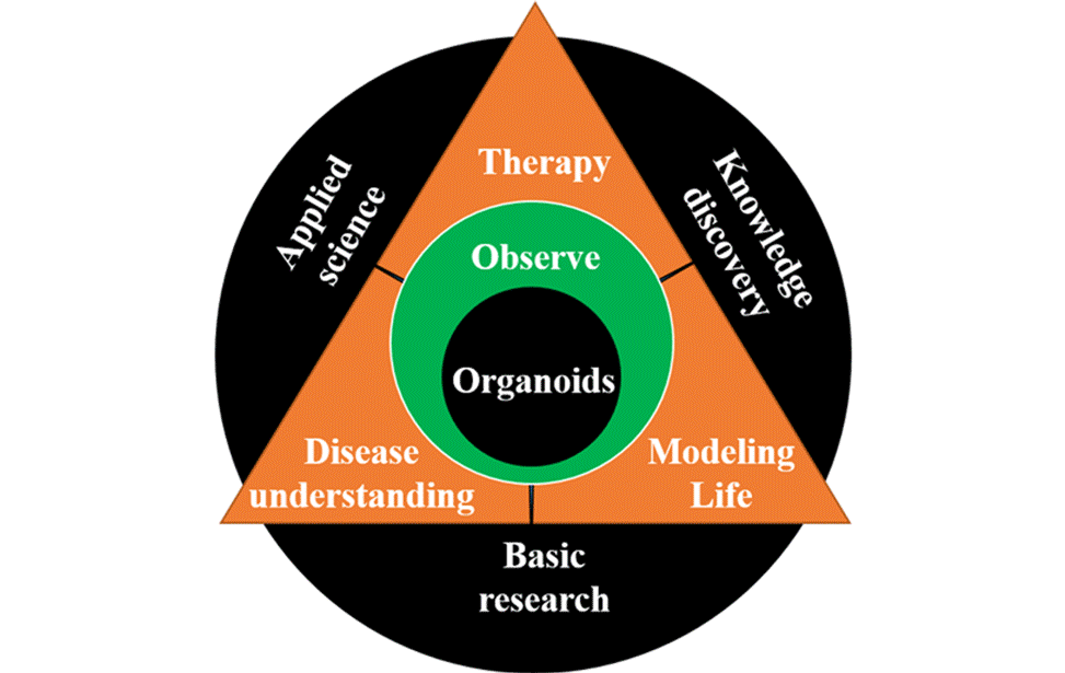
OrganVision, a technology proposal to image organoids real-time label-free, comprises 8 partners spread across 4 countries in Europe. OrganVision envisions to shift the paradigm for organoids from disease or drug-screening models to observable tissue micro-bio-environment to unravel a few key biomedical applications. We aspire to develop technology to unfold the real-time mechanism of an organoid system at sub-cellular and inter-cellular scales in cells and tissues, respectively.
Dipanjan Bhattacharya
Dipanjan Bhattacharya has multidisciplinary expertise in the field of microscopy, biophysics, and quantitative image analysis. He leads the Imaging Development Unit (IDU) of IFOM and is working on developing innovative imaging and image analysis technologies for cancer research and different biomedical applications. The Imaging Development Unit is particularly focussed on developing advanced imaging and image processing tools for processing for automated processing of large datatowards disease diagnosis and cure.
Dr. Bhattacharya did his Masters in Physics at the University of Kalyani, India. He then joined Raman Research Institute, Bangalore for his Ph.D. in Biophysics to pursue a collaborative project with National Center for Biological Sciences, TIFR, Bangalore, under the supervision of Dr. G.V. Shivashankar. His Ph.D thesis was aimed at understanding the role of histone dynamics and its functional implementation in live cells. During his Ph.D, he developed diverse microscopic techniques based on both force and fluorescence for quantitative biological applications.
For his postdoctoral work, he joined Prof. Paul Matsudaira’s lab at the Massachusetts Institute of Technology (MIT) in the Bio-Engineering dept. and worked in Whitehead Bioimaging Center. During this period, he enhanced his skills in Computational Biology and high-end Image Processing. Following this, he moved to Singapore and joined Singapore-MIT Alliance for Research and Technology Center (SMART), a newly developed campus of MIT in Singapore. At SMART he worked with Prof. Paul Matsudaira, Prof. George Barbastathis and Prof. Peter So, for developing diverse microscopic techniques, like Light-sheet microscopy (DSLM) with structured illumination, Volume Holographic Microscopy (MVH system), Confocal Phase Microscopy, Image Cytometry system, etc for various biomedical applications. He used these homebuilt imaging technologies to understand the mechano-biological cues of the convergence extension process in Zebrafish embryogenesis. Simultaneously, he also developed numerous computational tools for quantitative automated analysis of electron microscopy images. From 2012-17, he was jointly appointed as a research scientist in Mechanobiology Institute, Singapore and Singapore-MIT Alliance for Research and Technology Center (SMART).
In 2018, he moved to Milan to lead the Imaging development unit of IFOM. For developing innovating imaging and image analysis tools towards cancer biology and other biomedical applications.
In 2021, Dr. Bhattacharya is awarded FET Open grant from European Union’s Horizon-2020 along with his collaborators from four different European countries.