
July 10th- 21st, 2017 | IFOM, Milan - Italy
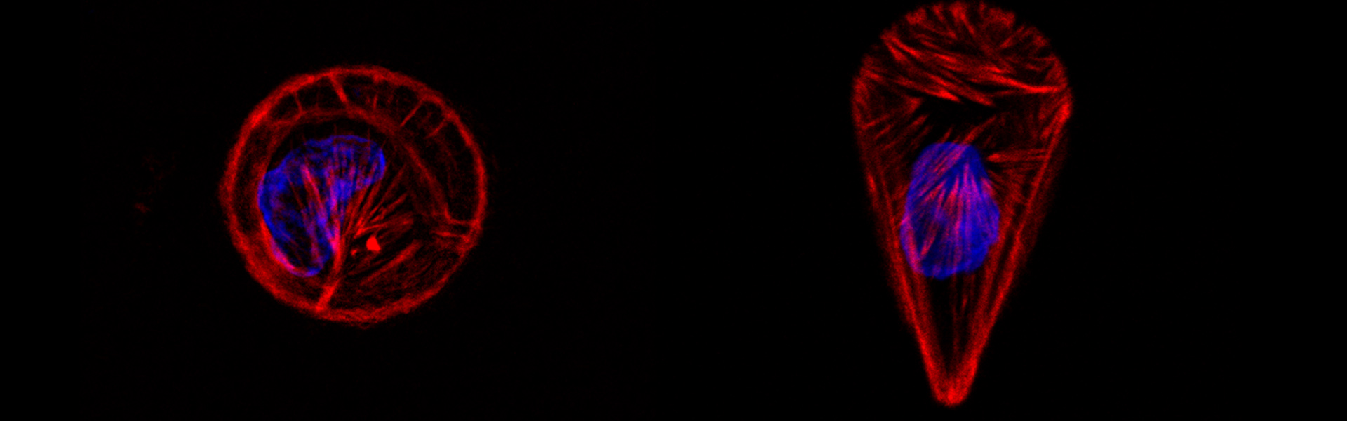
Cells in tissues are highly organized and have specific shapes that are strongly linked to their biological functions. This geometrical and spatial organization is essential for tissues homeostasis and can be partially recapitulated in 2D cell culture forcing cells to acquire a specific shape in vitro.
Micro-patterning of adhesive molecules is a technique used to control the cell geometry. With this method, adhesive molecules, like fibronectin, are patterned on a surface into well defined shapes which are surrounded by non-adhesive regions. Cells plated on this surface will then adjust their shape accordingly to the patterns as they can only adhere on the predetermined adhesive areas. By tuning size and shape of adhesive regions, it is possible to force single cells as well as multicellular complexes to acquire specific shapes or to move on lines or mazes.
Micro-patterning allows to partially mimic in 2D the complexity of the 3D cell micro-environment and to standardize the geometrical constrains of a cell population. It is a powerful and versatile technique employed often to study the organization of the cell cytoskeleton, cell division and cell migration. During the course students will learn a simple method to make micro-patterns and the basic knowledge of how to use microscopes to acquire images of fixed and living cells. They will quantitatively analyze their results and produce a report of their experiences.